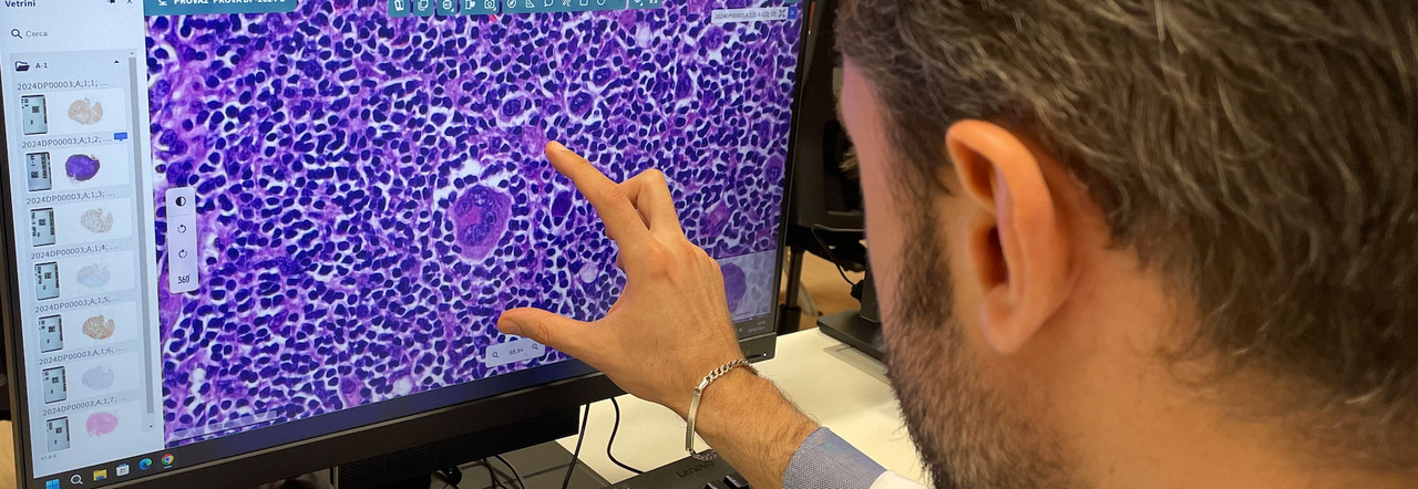
The operative unit of pathological anatomy of the Luigi Vanvitelli University Hospital becomes "digital" thanks to a unique pilot project in Campania. "The digitalization of the diagnostic process in pathological anatomy is a real revolution", explains Professor Renato Franco, director of the Operative Unit. "This change will have a huge positive impact on the diagnostic chain. Thanks to digital control, all the slides produced for diagnostic purposes from the lesions taken from patients are subjected to massive scans. In other words, we turn the slides into digital images that the pathologist can analyze from a latest generation Hd video-terminal".
The digitization project, strongly wanted by the general director Ferdinando Russo, with the support of the Magnificent Rector of the University of Campania "L. Vanvitelli" Gianfranco Nicoletti, leads from the start to a radical change for patients. Among the various implications "the sharing of individual images (and therefore of the case) suddenly becomes rapid, even more secure and multiple, for a collegial evaluation of the single diagnosis and a rapid sharing with experts in that specific pathology", emphasizes the general director Ferdinando Russo. In addition, thanks to the involvement of computer engineers, it is possible to use artificial intelligence programs for diagnostic support and the characterization of individual lesions.
Through the most modern digital infrastructures it is also possible to eliminate distances with the various presidia, which can ask for support in real time; even those that are very far from a department of pathological anatomy. "During surgical interventions - continues Professor Franco - it is possible that we find ourselves in front of something that was not predicted and that a rapid histological or cytological intraoperative examination is necessary to better replan the surgical-therapeutic path, in two words: an "extemporaneous examination". Thanks to digital microscopes and high-resolution scanners, it is possible to prepare a cyto-histological preparation during a surgical intervention and have it quickly examined remotely by an anatomical pathologist, instantly eliminating the distances that previously made this procedure impractical".
Also important is the possibility that Digital Pathology offers to share diagnostic material in Universities for the specialist training of doctors and future anatomical pathologists, to increase the available databases and information on individual pathologies. Meanwhile, already today (Wednesday 21 February) the Hospital Company and the University L. Vanvitelli have proposed a masterclass focused on the practical aspects of digital pathology operations, starting from the necessary infrastructure, the implementation of tools based on artificial intelligence and the financial aspects of managing a digital pathology operation. A meeting that allows participants to learn about new developments in the field of Digital Pathology and interact with other pathologists and researchers interested in promoting digital pathology in their institutions.

